June 2 is now World Interventional Pulmonology Day. Inside: ERS/ESGE/ESTS guidelines, C-Cryo-EBUS, two clinical cases, ROSE feedback, IPI Nairobi, deepdive Cologne and more.
Newsletter June 2026
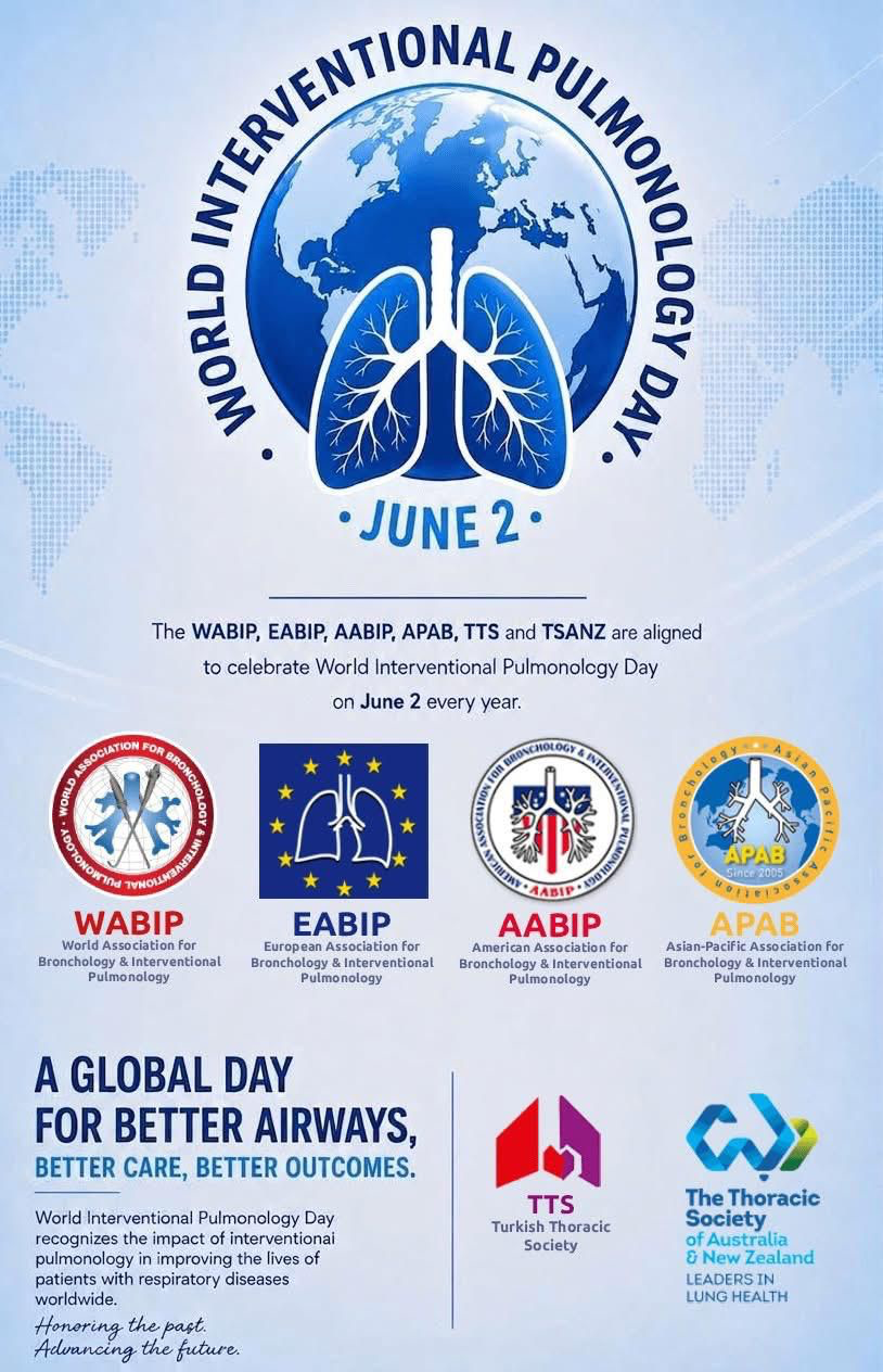
World Interventional Pulmonology Day
A new global milestone for our specialty: June 2 is now World Interventional Pulmonology Day.
Dear colleagues and friends,
We are delighted to share a historic announcement for the interventional pulmonology community worldwide: June 2 has officially been designated as World Interventional Pulmonology Day.
This landmark initiative is the result of an unprecedented collaboration between the World Association for Bronchology and Interventional Pulmonology (WABIP), the European Association for Bronchology and Interventional Pulmonology (EABIP), the American Association for Bronchology and Interventional Pulmonology (AABIP), the Asian-Pacific Association for Bronchology and Interventional Pulmonology (APAB), the Turkish Thoracic Society (TTS), and the Thoracic Society of Australia and New Zealand (TSANZ).
The date was chosen to commemorate the birthday of Gustav Killian, widely recognized as the pioneer of bronchoscopy, whose groundbreaking work laid the foundations of our specialty. By honoring his legacy, we also reaffirm our commitment to shaping the future of interventional pulmonology through innovation, education, research, and excellence in patient care.
World Interventional Pulmonology Day is more than a celebration: it is a global recognition of the profound impact our field has on the lives of patients with respiratory diseases. From advanced diagnostic procedures to minimally invasive therapeutic interventions, interventional pulmonology continues to transform the management of thoracic diseases and improve outcomes for millions of patients worldwide.
Under the shared theme “Better Airways, Better Care, Better Outcomes,” this annual observance will serve as a unifying platform for healthcare professionals, institutions, industry partners, and patient advocacy groups across all continents. It reflects a borderless commitment to advancing the science and practice of interventional pulmonology while ensuring equitable access to high-quality care.
As we celebrate the first World Interventional Pulmonology Day on June 2, we invite our global community to join us in recognizing the achievements of our specialty, inspiring future generations, and continuing the legacy of innovation that defines interventional pulmonology.
Honoring the past. Advancing the future.
🔬 Scientific & Clinical Updates
📖 Paper Highlight · ERS/ESGE/ESTS Guidelines
ERS/ESGE/ESTS Clinical Practice Guidelines on Endobronchial and Oesophageal Endosonography for the Diagnosis and Staging of Lung Cancer
Korevaar DA, Kovacevic B, Papadopoulou E, et al. Eur Respir J 2026 (Early View). DOI: 10.1183/13993003.00097-2026
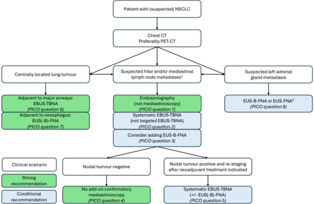
Figure 1 — Role of endosonography in patients with (suspected) NSCLC.
A new joint task force of the European Respiratory Society (ERS), the European Society of Gastrointestinal Endoscopy (ESGE) and the European Society of Thoracic Surgeons (ESTS) updates the 2015 guideline on endosonography in lung cancer. Twelve clinical questions were addressed using GRADE methodology, with consensus reached at an in-person task force meeting in Amsterdam (July 2025).
Key recommendations for (suspected) NSCLC: endosonography is recommended over mediastinoscopy for mediastinal nodal tissue staging; systematic staging is preferred over targeted staging; combined EBUS-TBNA plus EUS(-B)-FNA is preferred over EBUS-TBNA alone; add-on mediastinoscopy after a negative endosonography is not recommended; endosonography is suggested over mediastinoscopy for re-staging after induction therapy. 21G/22G TBNA needles remain the standard. EBUS-TBNA has a high suitability rate for PD-L1 assessment.
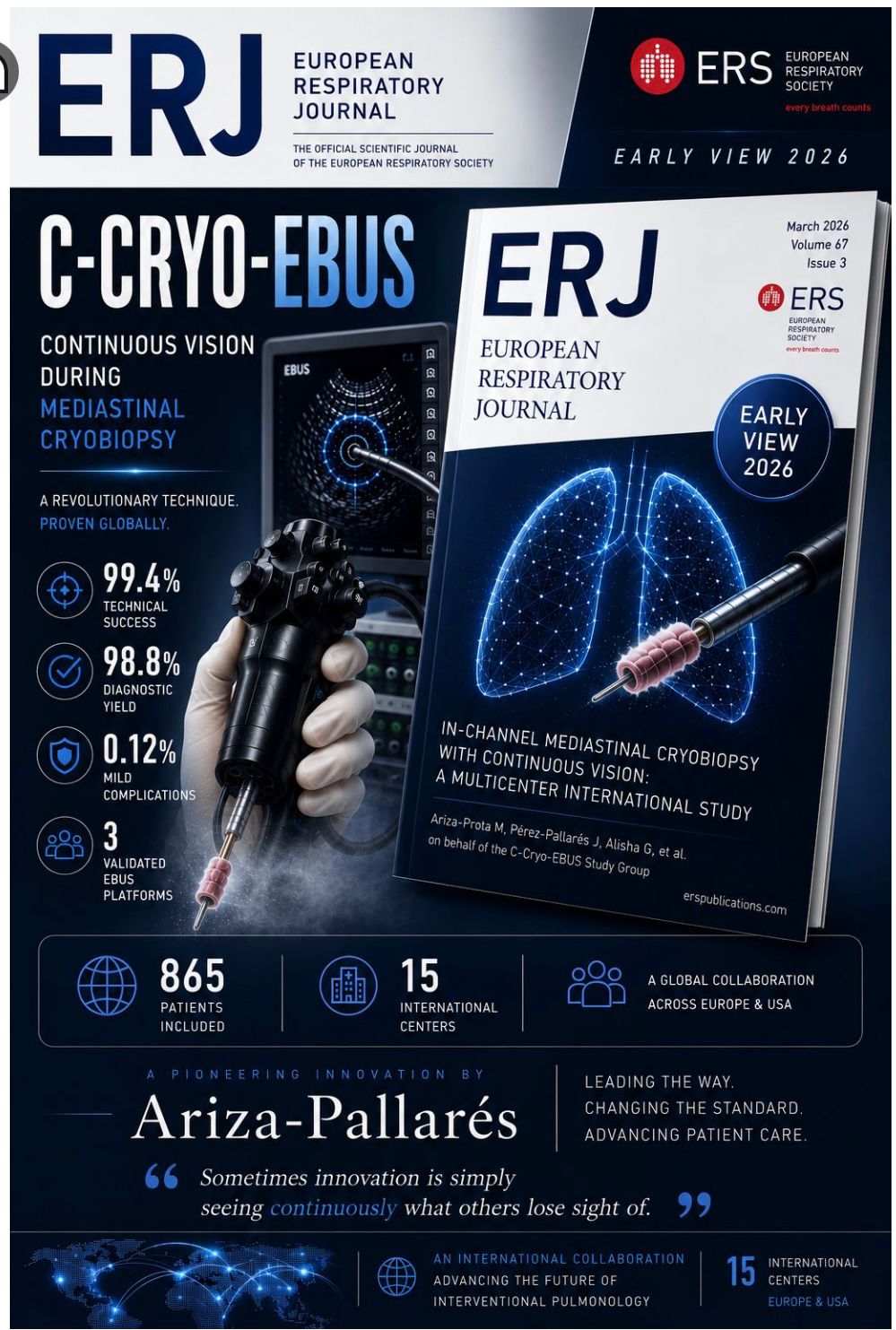
📖 Paper Highlight · International Multicenter Study
In-Channel Mediastinal Cryobiopsy (C-Cryo-EBUS): an International Multicenter Study of Feasibility, Safety, and Reproducibility
Ariza-Prota M, Pérez-Pallarés J, Alisha G, et al. Eur Respir J 2026 (Early View). DOI: 10.1183/13993003.00342-2026
An international, prospective, multicenter study conducted across 15 specialised interventional pulmonology units in Europe and the USA (March 2025 to January 2026) evaluated the feasibility, safety and reproducibility of in-channel mediastinal cryobiopsy (C-Cryo-EBUS) using a 1.1 mm cryoprobe, a standardised 3-second freezing protocol, and the Ariza-Pallárés tunneling technique. The cryoprobe is withdrawn through the working channel of the echobronchoscope without removing the scope, keeping continuous endoscopic visualisation.
Across 865 patients, technical success was 99.4% and overall diagnostic rate 98.8% (only 1.2% non-diagnostic). Sample adequacy for immunohistochemistry and molecular testing reached 95.3%. Mild complications were rare (0.12%). The technique was reproducible across all three commercially available EBUS platforms (Olympus, Fujifilm, Pentax) and particularly valuable in granulomatous disease and lymphoma subtyping.
How Fellowship Changed My Bronchoscopic Strategy: Endobronchial Hamartoma Debulking and Airway Recanalization
Dr. Dimas Bayu Firdaus, MD, Pulmonologist
Department of Pulmonology and Respiratory Medicine, Dr. M. Djamil Hospital / Universitas Andalas, Padang, Indonesia
Case summary
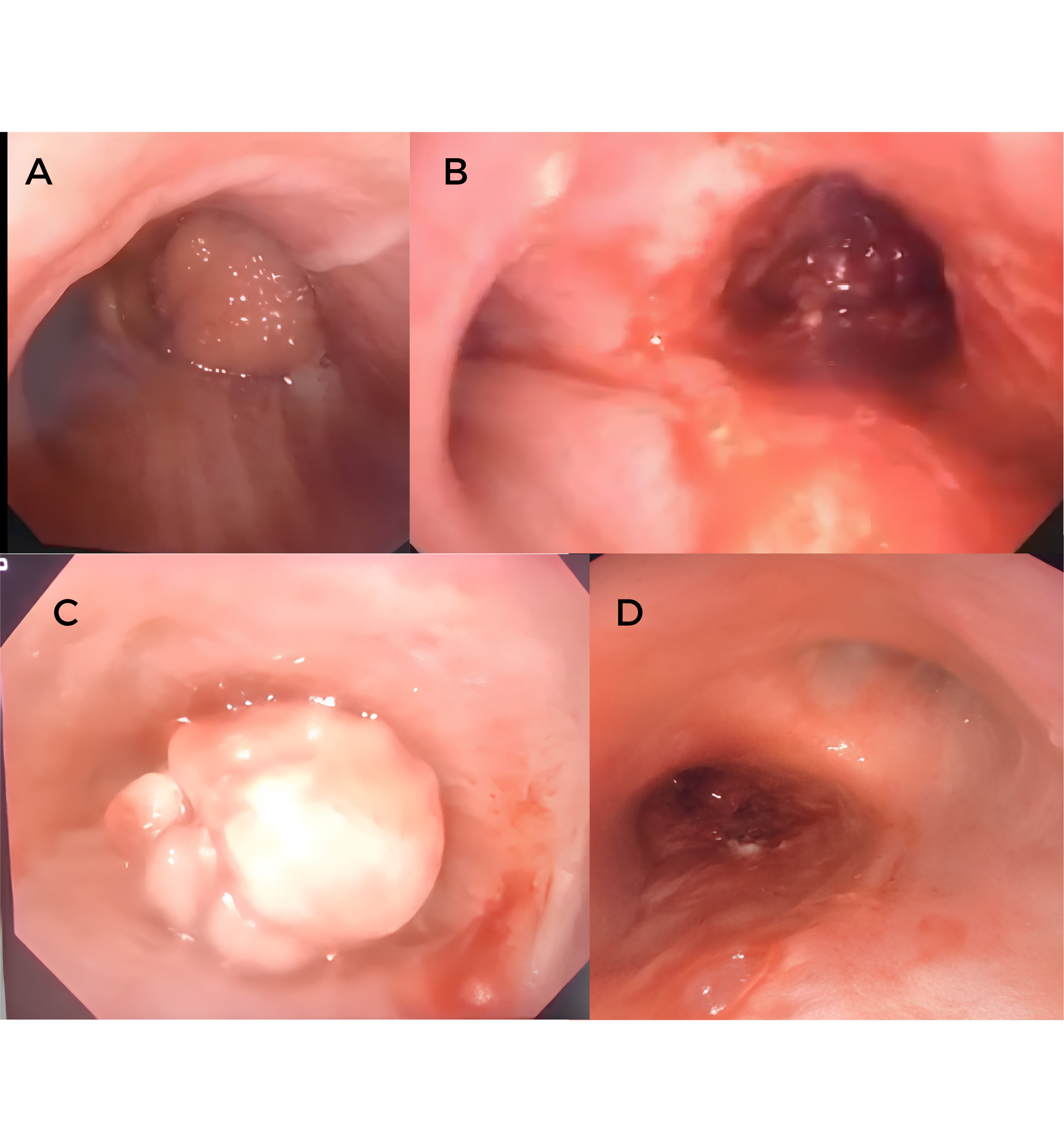
Staged bronchoscopic debulking of an endobronchial hamartoma causing right-sided airway obstruction. The case highlights how completing an interventional pulmonology fellowship reshaped the bronchoscopic strategy used to manage a complex central airway lesion, moving from a single-session approach to a planned, staged recanalization protocol.
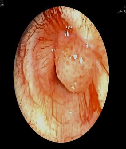
Fig. 1 — Bronchoscopic views during the staged debulking: the obstructing endobronchial mass, intra-procedural appearance, and progressive restoration of the right-sided airway lumen.
Imaging and procedural course
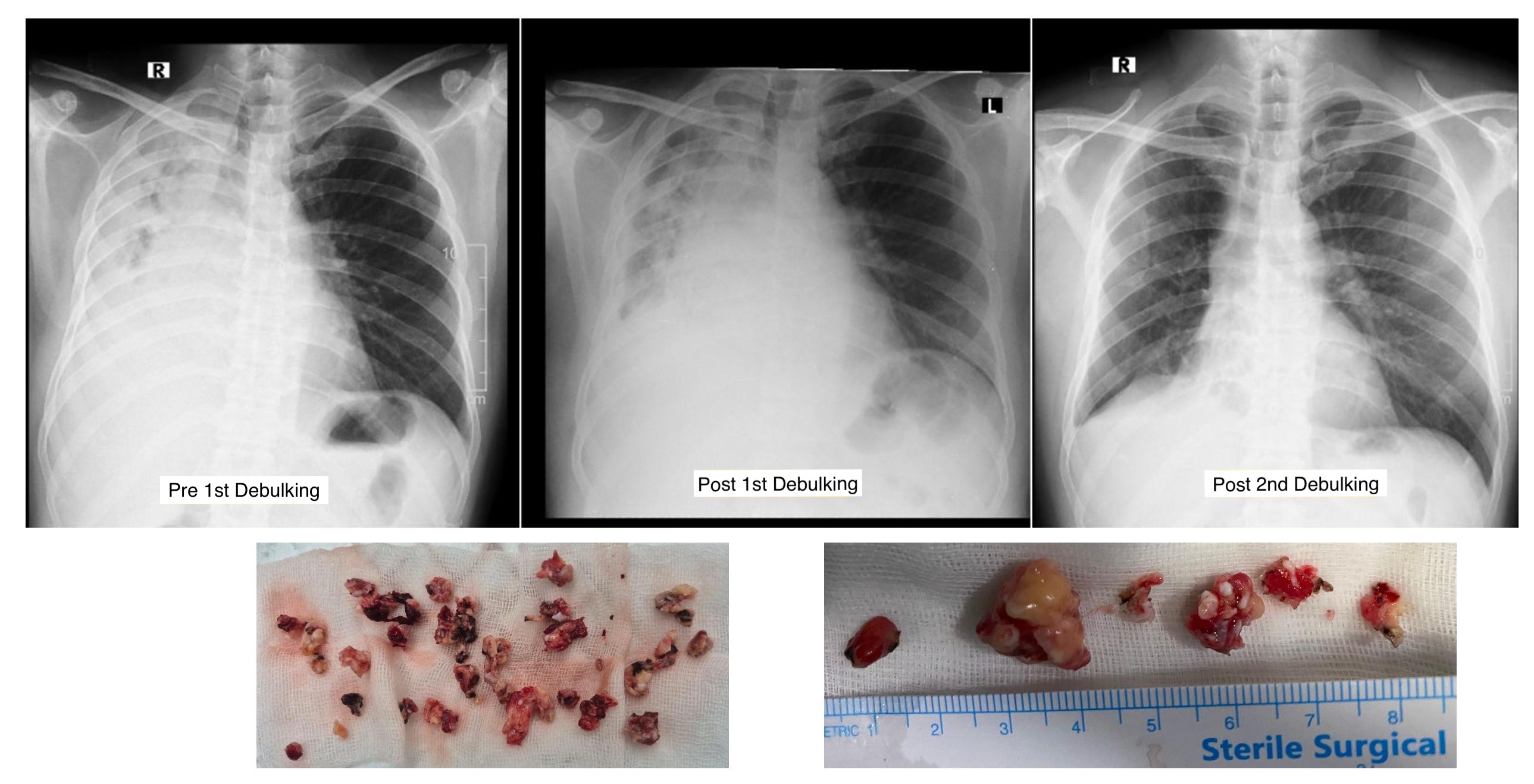
Baseline chest X-ray showed complete opacification of the right lung due to the obstructing endobronchial lesion. A first bronchoscopic debulking session restored partial airway patency and re-aeration of the right lung. A second debulking session, performed after reassessment of residual disease, achieved near-complete recanalization and recovery of normal lung volumes on follow-up imaging. Macroscopic examination of the retrieved fragments was consistent with an endobronchial hamartoma.
Fig. 2 — Top row: serial chest X-rays showing Pre 1st Debulking (complete right lung opacification), Post 1st Debulking (partial re-aeration) and Post 2nd Debulking (near-complete re-aeration). Bottom row: macroscopic specimens of the hamartoma fragments retrieved during the two debulking sessions.
Final diagnosis
Endobronchial hamartoma.
🔍 Procedure Spotlight
Microlaryngeal Tube assisted bronchoscopic management of central airway obstruction: A novel technique
Dr. Viswesvaran Balasubramanian, MD, DM & Dr. Pothireddy Manisha Reddy, MD
Yashoda Hospitals, Somajiguda, Hyderabad, India
Case presentation
A 26-year-old female presented with progressive dyspnea and refractory cough. On examination, the patient was tachypneic. Initial evaluation, including imaging and flexible bronchoscopy, revealed a near-complete obstruction of the central airway by an endotracheal mass. Given the critical nature of the airway compromise, she was referred from a tertiary care center approximately 600 km away for urgent bronchoscopic intervention.
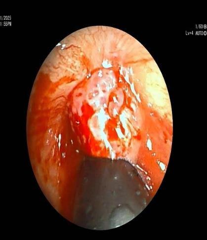
Fig. 1 — Bronchoscopic image of vascular tracheal mass causing near-complete occlusion.
Bronchoscopic intervention
A multidisciplinary team approach was adopted and the procedure was performed using awake bronchoscopic intubation with a microlaryngeal tube (MLT) to maintain ventilation and secure distal airway access. Careful and meticulous placement of the MLT was ensured to avoid trauma to the vascular tumor. Following airway stabilization, bronchoscopic tumor debulking was carried out using a multimodality approach: electrosurgical snaring was initially performed to reduce the bulk of the lesion, followed by cryoextraction to remove residual tumor tissue. After achieving adequate airway clearance, a self-expanding metallic stent (SEMS) was deployed to maintain airway patency.
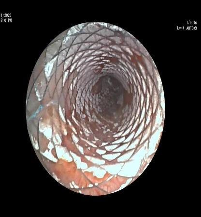
Fig. 2 — Microlaryngeal tube placement distal to the mass.
Fig. 3 — Post tracheal SEMS placement.
Outcome
The procedure resulted in successful recanalisation of the airway with immediate improvement in ventilation. The patient was extubated on the table and remained hemodynamically stable throughout. She demonstrated rapid clinical recovery and was discharged on the same day in an ambulatory condition. Histopathological examination of the resected tissue revealed high-grade Mucoepidermoid carcinoma with mediastinal extension, rendering the tumor surgically inoperable. The patient was subsequently planned for definitive chemoradiation therapy.
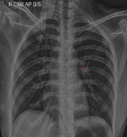
Fig. 4 — Post procedure chest X-ray.
🎥 Procedure video
Video. MLT-assisted bronchoscopic management of central airway obstruction by a high-grade mucoepidermoid carcinoma.
🤝 Community & Networking
Expanding Global Access to Interventional Pulmonology Training
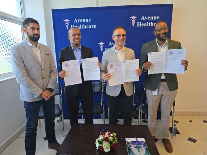
Launch of the 2nd WABIP Interventional Pulmonology Institute (IPI Nairobi)
Avenue Hospital, Nairobi, Kenya
📆 Fellowship programme starts January 2027
A joint WABIP & Avenue Hospital initiative
The World Association for Bronchology and Interventional Pulmonology (WABIP) has officially inaugurated its second Interventional Pulmonology Institute (IPI) in Nairobi, in partnership with Avenue Hospital. Building on the success of the pioneering IPI Istanbul, which has trained eight fellows annually from around the world, IPI Nairobi will open its fellowship training programme in January 2027.
The programme will be led by Prof. Ali Musani (President-elect WABIP and Chair of the IPI programmes globally), alongside Dr. Naveed Merali (Director of IPI Nairobi and a graduate of the IPI Istanbul programme), supported by an international faculty. Trainees will gain comprehensive theoretical knowledge and hands-on experience in advanced diagnostic and therapeutic bronchoscopy within a high-volume, state-of-the-art training centre.
Across Africa, fewer than five centres in the East currently offer key procedures such as EBUS or rigid bronchoscopy, and only one centre in East and Central Africa performs complex interventions such as navigation bronchoscopy or airway stenting. IPI Nairobi addresses this gap and is designed to serve physicians from across Africa and from developing countries worldwide, expanding equitable access to high-quality respiratory care.
Pleural Effusion Quantification & Assessment: An International Survey
The Imaging Study Group of the Italian Respiratory Society (SIP-IRS), led by Dr. Guido Marchi (Pisa University Hospital), is inviting clinicians worldwide to participate in an international survey on “Optimal pleural effusion quantification and assessment”.
The survey has a dual objective:
• Characterise current clinical practices and perceived challenges in pleural effusion quantification.
• Assess clinicians' familiarity with, and expectations regarding, emerging technologies such as handheld ultrasound devices and AI-based decision support tools.
The survey is anonymous, takes only a few minutes to complete, and is open to all physicians involved in the management of pleural effusion, regardless of specialty or clinical setting.
Your participation contributes to identifying priority areas for the standardisation of clinical practice and to guiding future research in this field.
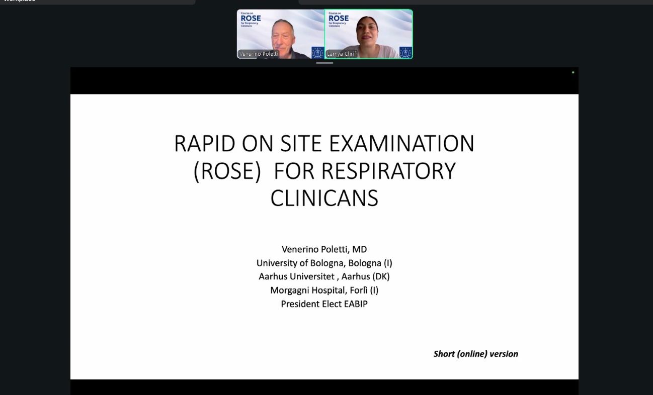
Course on Rapid On-Site Examination (ROSE) for Respiratory Clinicians
On 21 and 22 May 2026, the EABIP hosted a highly successful live webinar dedicated to Rapid On-Site Examination (ROSE) for respiratory clinicians. The course was delivered by Prof. Venerino Poletti (University of Bologna, Aarhus Universitet, Morgagni Hospital, Forlì - President Elect EABIP) and moderated by Dr. Lamya Chrif Morand (Director of Communication & Public Affairs, EABIP).
Opening of the ROSE webinar with Prof. Venerino Poletti and Dr. Lamya Chrif Morand.
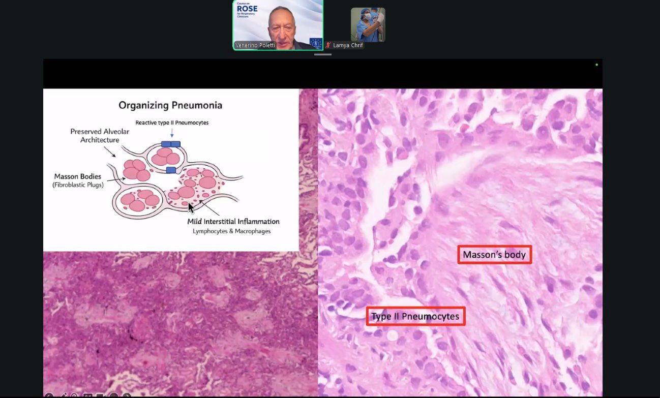
The course attracted a large international audience and generated active discussions throughout both sessions. Participants engaged with practical cytology examples, learned how to integrate ROSE into routine bronchoscopy workflows, and explored real-time decision-making for diffuse parenchymal lung disease, granulomatous disorders and lymphoproliferative entities.
Course slide: organising pneumonia, Masson's body and type II pneumocytes, an example of the cytopathology material discussed live.
Sincere thanks to Prof. Poletti for sharing his expertise and delivering such an insightful and inspiring course, and to Dr. Lamya Chrif Morand for the excellent moderation that made the session engaging and impactful.
Looking forward to many more impactful learning opportunities together. #ROSE #Pulmonology #Bronchoscopy #MedicalEducation
📅 Upcoming Events
4th Global Conference on Robotic Assisted Bronchoscopy
📍 Brescia, Italy
📆 18–19 June 2026
RAB 2026 is back in Italy! The 4th Global Conference on Robotic Assisted Bronchoscopy will return to its birthplace in Brescia, bringing together leading experts in the field.
Masterclass for bronchoscopic lung diagnostics and therapy
deepdive Bronchoscopy Masterclass — 2nd edition
Lead by
Priv.-Doz. Dr. med. Filiz Özkan
Universitätsmedizin Essen, Ruhrlandklinik
Dr. med. Jane Winantea
Universitätsmedizin Essen, Ruhrlandklinik
Dr. med. Lea Marie Schotten
Helios Klinikum Bonn / Rhein-Sieg, Ruhrlandklinik
Steigenberger Hotel Cologne, Germany
📆 17–19 September 2026
A practice-oriented, interactive masterclass on diagnostic and therapeutic bronchoscopy for lung cancer. Live demonstrations, hands-on workshop rotations, and lectures from leading European and international faculty cover EBUS-guided cryobiopsy, robotic navigation, ICG coils, recanalisation and stenting, microwave ablation, and the latest updates in SCLC and NSCLC therapy.
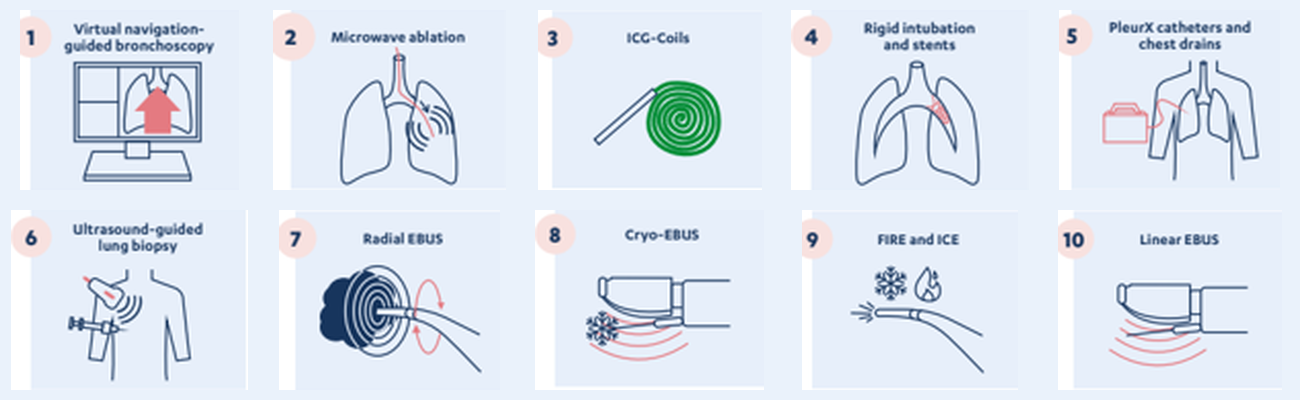
Hands-on workshop stations
Ten hands-on workshop stations covering navigation, ablation, stenting, pleural procedures, and the full EBUS toolbox.
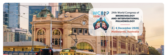
24th World Congress of Bronchology and Interventional Pulmonology
📍 Melbourne, Australia
📆 3–6 December 2026
Hosted by TSANZ & WABIP
The premier international meeting in bronchology and interventional pulmonology returns for its 24th edition. Join colleagues from around the world for cutting-edge science, hands-on workshops, and networking opportunities.
Abstract submissions are now open, don’t miss the opportunity to present your work on the world stage.
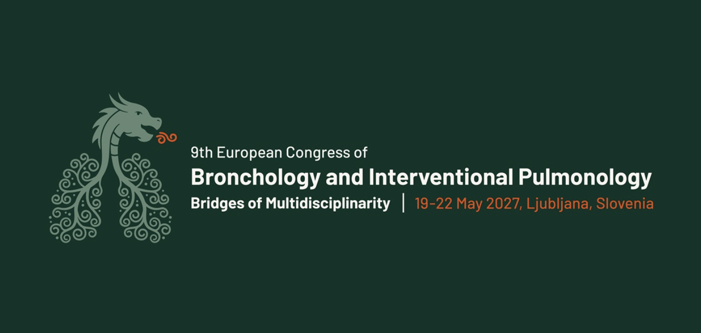
9th European Congress of Bronchology and Interventional Pulmonology
📍 Ljubljana, Slovenia
📆 19–22 May 2027
“Bridges of Multidisciplinarity”
The 9th ECBIP will gather European and international experts to advance the frontiers of bronchology and interventional pulmonology through multidisciplinary collaboration.
Save the date, more details on registration and abstract submission coming soon.
• Share initiatives and collaborations
• Submit educational material
• Promote national and international projects
Together, we continue to build a strong, united, and forward-looking interventional pulmonology network.
Share your best moments!
You are invited to share your best bronchoscopic images and videos of airway and/or pleural abnormalities. Selected submissions will be featured in upcoming EABIP Newsletter issues and shared across our social media channels (LinkedIn, Instagram, Facebook).